1. Internal and External Anatomy of the Kidney
External Anatomy
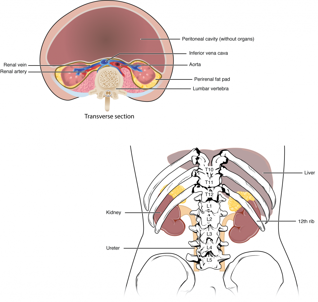
The paired kidneys lie on either side of the spine in the retroperitoneal space between the parietal peritoneum and the posterior abdominal wall, well protected by muscle, fat, and ribs. The left kidney is located at about the T12 to L3 vertebrae, whereas the right is lower due to slight displacement by the liver. Upper portions of the kidneys are somewhat protected by the eleventh and twelfth ribs (Figure 1.1). Each kidney weighs about 125–175 g in males and 115–155 g in females. They are about 11–14 cm in length, 6 cm wide, and 4 cm thick, and are directly covered by a fibrous capsule composed of dense, irregular connective tissue that helps to hold their shape and protect them. This capsule is covered by a shock-absorbing layer of adipose tissue called the renal fat pad, which in turn is encompassed by a tough renal fascia. The fascia and, to a lesser extent, the overlying peritoneum serve to firmly anchor the kidneys to the posterior abdominal wall in a retroperitoneal position.
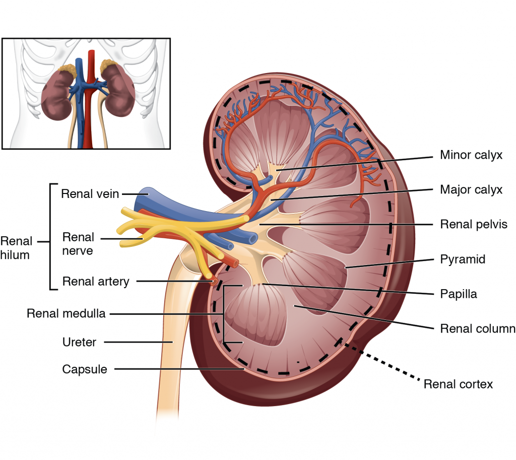
Each kidney looks like the kidney bean and the renal hilum is the entry and exit site for structures servicing the kidneys: vessels, nerves, lymphatics, and ureters. The medial-facing hila are tucked into the convex indentation of the kidney.
Internal Anatomy
A frontal section through the kidney reveals an outer region called the renal cortex and an inner region called the renal medulla (Figure 1.2). In the medulla, 5-8 renal pyramids are separated by connective tissue renal columns. Each pyramid creates urine and terminates into a renal papilla. Each renal papilla drains into a collecting pool called a minor calyx; several minor calyces connect to form a major calyx; all major calyces connect to the single renal pelvis which connects to the ureter.
Blood Supply of the Kidney & Nephrons
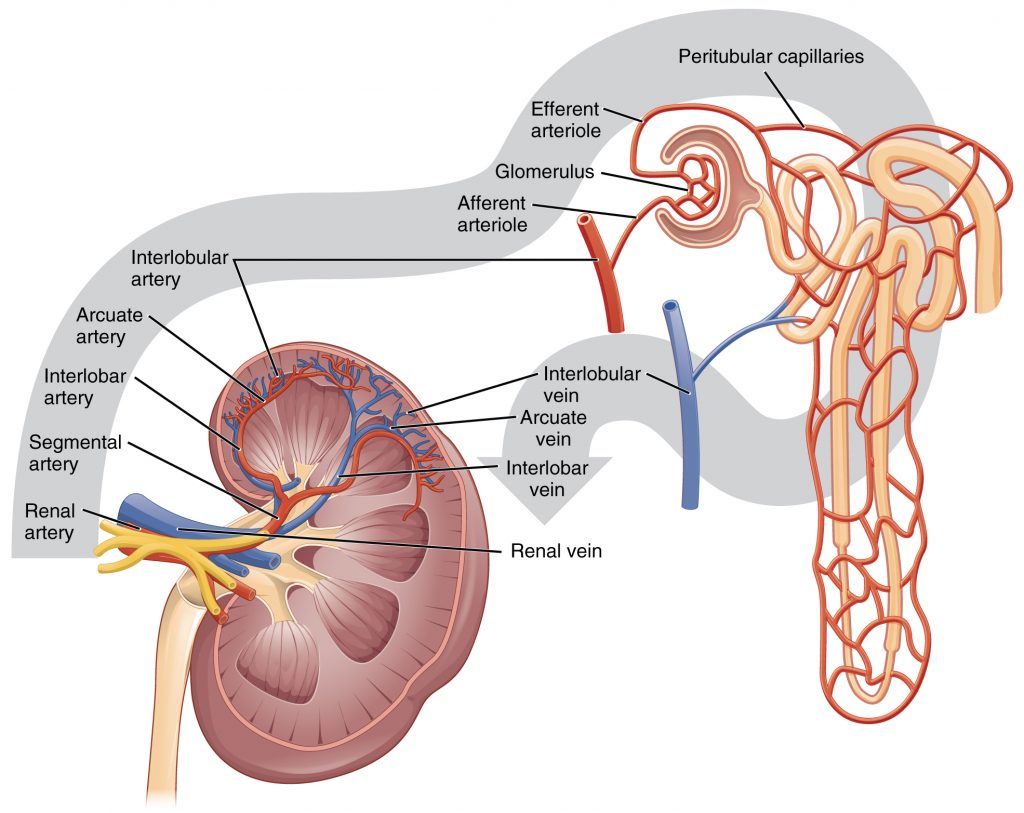
The kidneys are well vascularized and receive about 25 percent of the cardiac output at rest. Blood enters the kidney via the paired renal arteries that form directly from the descending aorta and each enters the kidney at the renal hila. Once in the kidney, each renal artery first divides into segmental arteries, followed by further branching to form interlobar arteries that pass through the renal columns to reach the cortex (Figure 1.3). The interlobar arteries, in turn, branch into arcuate arteries, cortical radiate arteries, and then into afferent arterioles. The afferent arterioles deliver blood into a modified capillary bed called the glomerulus which is a component of the “functional unit” of the kidney called the nephron. There are about 1.3 million nephrons in each kidney and they function to filter the blood. Once the nephrons have filtered the blood, renal veins return blood directly to the inferior vena cava. A portal system is formed when the blood flows from the glomerulus to the efferent arteriole through a second capillary bed, the peritubular capillaries (and vasa recta), surrounding the proximal and distal convoluted tubules and the loop of Henle. Most water and solutes are recovered by this second capillary bed. This filtrate is processed and finally gathered by collecting ducts that drain into the minor calyces, which merge to form major calyces; the filtrate then proceeds to the renal pelvis and finally the ureters.